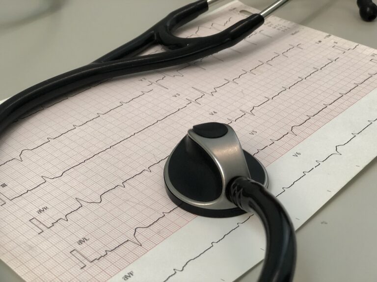
Electrocardiogram (ECG): what it is for and how it is performed
The electrocardiogram is a diagnostic test. Using the electrocardiograph, it records the rhythm and electrical activity of the heart.

The electrocardiogram is a diagnostic test. The electrocardiograph records the rhythm and electrical activity of the heart in a non-invasive way. The recording of the heart rhythm is shown in a tracing. By looking at the tracing (called an ECG), the cardiologist can assess the health of the heart.
Electrocardiogram: what it detects
Various cardiac abnormalities can be detected from the electrocardiogram. In fact, the ECG tracing can show different heart conditions that may indicate:
- Atrial abnormalities
- Abnormalities of the ventricle of the heart
- Arrhythmias (changes in the heart rhythm)
- Correct functioning of an implanted cardioverter defibrillator
- Correct functioning of the pacemaker
- Coronary damage
- Signs of a previous or current myocardial infarction
- Other heart disease conditions
Types of electrocardiogram
For a patient, the ECG can be performed in different ways.
- At rest
- 24-hour dynamic ECG (Holter)
- Under stress
The test at rest is also called the baseline electrocardiogram.
Prevention
The electrocardiogram is a very simple diagnostic tool that everyone, men and women, should use. Especially after age 40 it has considerable importance. In particular for people who suffer from:
- Diabetes
- High blood pressure
- Kidney disease
- Conditions of various kinds
Certain categories of people should also have a periodic follow-up electrocardiogram:
- People undergoing diagnostic tests with a contrast agent
- Healthy people who have periodic checkups
- Athletes
The ECG is the first cardiology diagnostic test to be done before all the others, and it must also come before a cardiac ultrasound, a cardiac CT scan, etc., etc…
Having an ECG: what to wear
The electrocardiogram is a non-invasive diagnostic test. A key point is that the patient should be relaxed when the test is performed at rest. The wrists, ankles and chest need to be uncovered. In addition, watches, bracelets, rings and necklaces must be removed. Women must have the ECG without wearing a bra, so that the electrodes can be placed on the chest. It is preferable not to wear an undershirt during the test, and if there is excess hair on the chest it is advisable to remove it.
ECG in pregnancy
The electrocardiogram is one of the tests requested for pregnant women. The doctor usually prescribes it around the 37th week of gestation if delivery is expected at the 40th. If a cesarean delivery has already been planned by the gynecologist, the electrocardiogram can also be done before the 37th week. It is essential, however, that it is always the doctor who requests it. The specialist does not always prescribe the test, however. In fact, the ECG is also requested for pregnant women because of cardiovascular problems. To women over 35 years of age, or to former smokers. It is also very useful for anyone who has not had this diagnostic test in the last 3 years. A slight tachycardia, leading to an increase of about 15 beats per minute, is considered normal in pregnant women, due to the development of the fetus. In addition, tachycardia in pregnancy can also be due to factors such as:
- Anemia
- Anxiety
- Undiagnosed heart disease
- Thyroid dysfunction
Normal electrocardiogram
An ECG is normal when the electrocardiograph tracing has very specific characteristics. In fact, a person with no heart problems has a tracing that shows 5 typical waves. These are lines identified by the letters P, Q, R, S, T. The cardiac cycle that the cardiologist observes is assessed on the PQRST complex.
The function of the electrocardiograph
Through the electrodes placed on the patient’s body, the electrocardiograph records cardiac function, translating it onto a monitor and/or onto a roll of graph paper. The graph that the cardiologist reads is the tracing that shows waves, that is, lines that define the electrical activity of the heart.
Nefrocenter Cardio
Nefrocenter Cardio is a high-quality facility. It is dedicated to assessing the health of the heart and to detecting early signs of heart damage. The staff of Nefrocenter Cardio are very highly qualified professionals. They are able not only to read an electrocardiogram optimally, but also to carry out an accurate and meticulous cardiology examination which, combined with the interpretation of the electrocardiographic tracing, provides a service of high clinical value for the prevention and treatment of cardiovascular diseases.